Leg Muscle Diagram Anterior / Muscles of the Posterior Leg - Attachments - Actions ... : The tibialis anterior (tibialis anticus) is situated on the lateral side of the tibia;
byDawn Guerra•
0
Leg Muscle Diagram Anterior / Muscles of the Posterior Leg - Attachments - Actions ... : The tibialis anterior (tibialis anticus) is situated on the lateral side of the tibia;. The extensor hallucis longus is a thin muscle, situated within the tibialis anterior and the extensor digitorum longus. Quad leg muscles leg muscles diagram leg muscles anatomy muscle diagram muscle anatomy quad anatomy foot reflexology workout posters science biology. Muscles, connected to bones or internal organs and blood vessels, are in charge for movement. The human leg, in the general word sense, is the entire lower limb of the human body, including the foot, thigh and even the hip or gluteal region. Muscles within this compartment primarily produce ankle dorsiflexion and toe extension.
Extensor muscle group of lower leg. Detailed anterior, lateral and posterior views.men sports fitness training. The extensor hallucis longus is a thin muscle, situated within the tibialis anterior and the extensor digitorum longus. However, the definition in human anatomy refers only to the section of the lower limb extending from the knee to the ankle. This muscle runs from the tibia to the big toe.
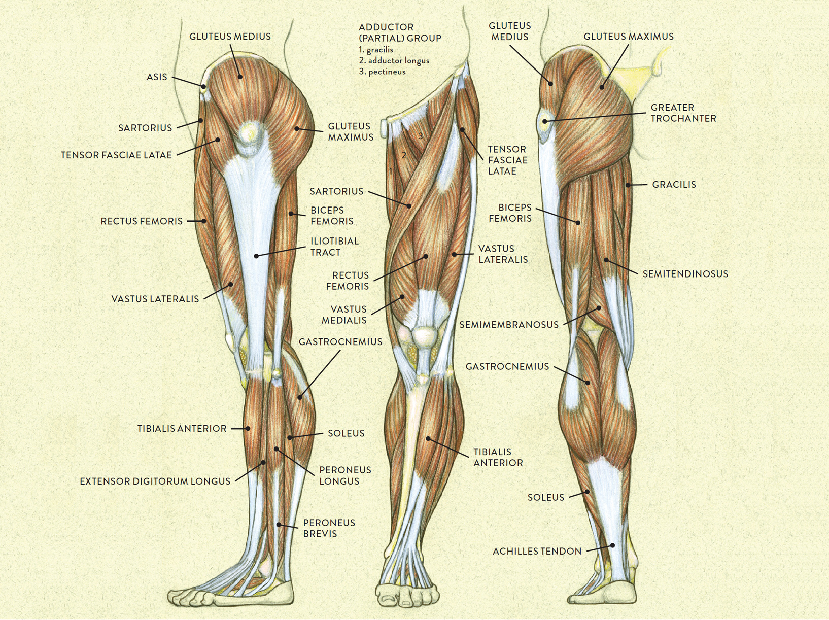
Lower Extremity from classroom.sdmesa.edu The accompanying muscle diagram reveals the position of the muscles of the lower legs in this pose. The human leg, in the general word sense, is the entire lower limb of the human body, including the foot, thigh and even the hip or gluteal region. Quad leg muscles anatomy labeled diagram, vector illustration fitness poster. The muscles of the leg may be divided into three groups: Quad leg muscles leg muscles diagram leg muscles anatomy muscle diagram muscle anatomy quad anatomy foot reflexology workout posters science biology. This muscle originates on the anterior lateral shaft of the femur and distal lines aspera and inserts onto the tibial tuberosity via the patellar ligament/quadriceps tendon. Extensor muscle group of lower leg. It contains muscles that produce dorsiflexion and participate in inversion and eversion of the foot, as well as vascular and nervous elements including the anterior tibial artery and veins, and the deep fibular nerve.
The extensor hallucis longus belongs to the anterior #muscles of the lower leg.
Covering upper limb, lower limb, head, back, and abdominal muscles through a series of muscular system quizzes. Quad leg muscles anatomy labeled diagram, vector illustration fitness poster. The extensor digitorum longus and extensor hallucis longus also extend the toes. A muscle along the outside of the leg that bends the foot out at the ankle. Gastrocnemius soleus peroneus longus tibialis anterior extensor digitorum longus extensor hallucis longus flexor digitorum. Produce wrist and/or finger flexion. The muscles of the leg may be divided into three groups: Vector illustration informative medical scheme. Each of these muscles is a discrete organ constructed of skeletal muscle tissue, blood vessels, tendons, and nerves. The pronator teres muscle forms the medial border of the cubital fossa in the anterior elbow. A complete list of muscular system quizzes; What is the origin of tibialis anterior? The extensor hallucis longus belongs to the anterior #muscles of the lower leg.
Detailed anterior, lateral and posterior views.men sports fitness training. It originates from the proximal portion of the leg, precisely. Quad leg muscles leg muscles diagram leg muscles anatomy muscle diagram muscle anatomy quad anatomy foot reflexology workout posters science biology. There are four muscles in the anterior compartment of the leg. Each of these muscles is a discrete organ constructed of skeletal muscle tissue, blood vessels, tendons, and nerves.
Muscles of the Leg and Foot - Classic Human Anatomy in ... from schoolbag.info A muscle along the outside of the leg that bends the foot out at the ankle. Human leg muscles diagram leg muscle chart gosutalentrankco. However, the definition in human anatomy refers only to the section of the lower limb extending from the knee to the ankle. Muscles of the anterior compartment of the forearm. Extensor muscle group of lower leg. The muscles of the anterior leg are located within the anterior compartment of the leg. It originates from the proximal portion of the leg, precisely. This muscle runs from the tibia to the big toe.
The deep muscles that impact leg movement are generally smaller that those that are directly involved in flexing the knee.
This is a preview of the anterior and lateral muscles of the leg anatomy video tutorial. The leg is separated into anterior, lateral, superficial posterior and deep posterior compartments by intermuscular septa and surrounded by the deep fascia of the leg. In this image, you will find anterior superior iliac spine, iliopsoas, tensor fasciae latae, sartorius, iliotibial tract, rectus femoris, vastus lateralis, patella, patellar ligament, peroneus longus, extensor digitorum longus, inguinal ligament, femoral artery, femoral vein, superficial inguinal ring, pectineus. The extensor hallucis longus is a thin muscle, situated within the tibialis anterior and the extensor digitorum longus. Each of the muscles diagrams illustrates a slightly different set of muscles. Lateral condyle & superior half of tibia. Medial cuneiform & 1st metatarsal. Each of these muscles is a discrete organ constructed of skeletal muscle tissue, blood vessels, tendons, and nerves. It is thick and fleshy above, tendinous below. The anterior muscles are a group of extensors at the lower leg. Pin our muscle facts and master #anatomy today. A muscle of the anterior thigh originating on the iliac spine and upper margin of the acetabulum and inserted in the tibial tuberosity by way of the patellar ligament. Muscles of the anterior compartment of the forearm.
Vector illustration informative medical scheme. Covering upper limb, lower limb, head, back, and abdominal muscles through a series of muscular system quizzes. Extensor muscle group of lower leg. Pin our muscle facts and master #anatomy today. Detailed anterior, lateral and posterior views.men sports fitness training.
Female Anterior Leg Muscles Labeled Educational Chart inch ... from s3.amazonaws.com In this image, you will find anterior superior iliac spine, iliopsoas, tensor fasciae latae, sartorius, iliotibial tract, rectus femoris, vastus lateralis, patella, patellar ligament, peroneus longus, extensor digitorum longus, inguinal ligament, femoral artery, femoral vein, superficial inguinal ring, pectineus. Produce wrist and/or finger flexion. Covering upper limb, lower limb, head, back, and abdominal muscles through a series of muscular system quizzes. Anterior muscles in the body. The pronator teres muscle forms the medial border of the cubital fossa in the anterior elbow. This muscle originates on the anterior lateral shaft of the femur and distal lines aspera and inserts onto the tibial tuberosity via the patellar ligament/quadriceps tendon. Almost every movement in the body is the outcome of muscle contraction. Where does tibialis anterior insert?
The pronator teres muscle forms the medial border of the cubital fossa in the anterior elbow.
It originates from the proximal portion of the leg, precisely. Pin our muscle facts and master #anatomy today. Medial cuneiform & 1st metatarsal. The extensor hallucis longus belongs to the anterior #muscles of the lower leg. Produce wrist and/or finger flexion. Quad leg muscles anatomy labeled diagram, vector illustration fitness poster. The accompanying muscle diagram reveals the position of the muscles of the lower legs in this pose. Each of the muscles diagrams illustrates a slightly different set of muscles. In this image, you will find anterior superior iliac spine, iliopsoas, tensor fasciae latae, sartorius, iliotibial tract, rectus femoris, vastus lateralis, patella, patellar ligament, peroneus longus, extensor digitorum longus, inguinal ligament, femoral artery, femoral vein, superficial inguinal ring, pectineus. Muscles of the anterior compartment of the forearm. The deep muscles that impact leg movement are generally smaller that those that are directly involved in flexing the knee. There are four muscles in the anterior compartment of the leg. The extensor hallucis longus belongs to the anterior #muscles of the lower leg.
This is a preview of the anterior and lateral muscles of the leg anatomy video tutorial leg muscle diagram. Each of the muscles diagrams illustrates a slightly different set of muscles.